Heartland Medical Specialties, Inc.
Chison Ultra Sound for GYN Features
EBit 50
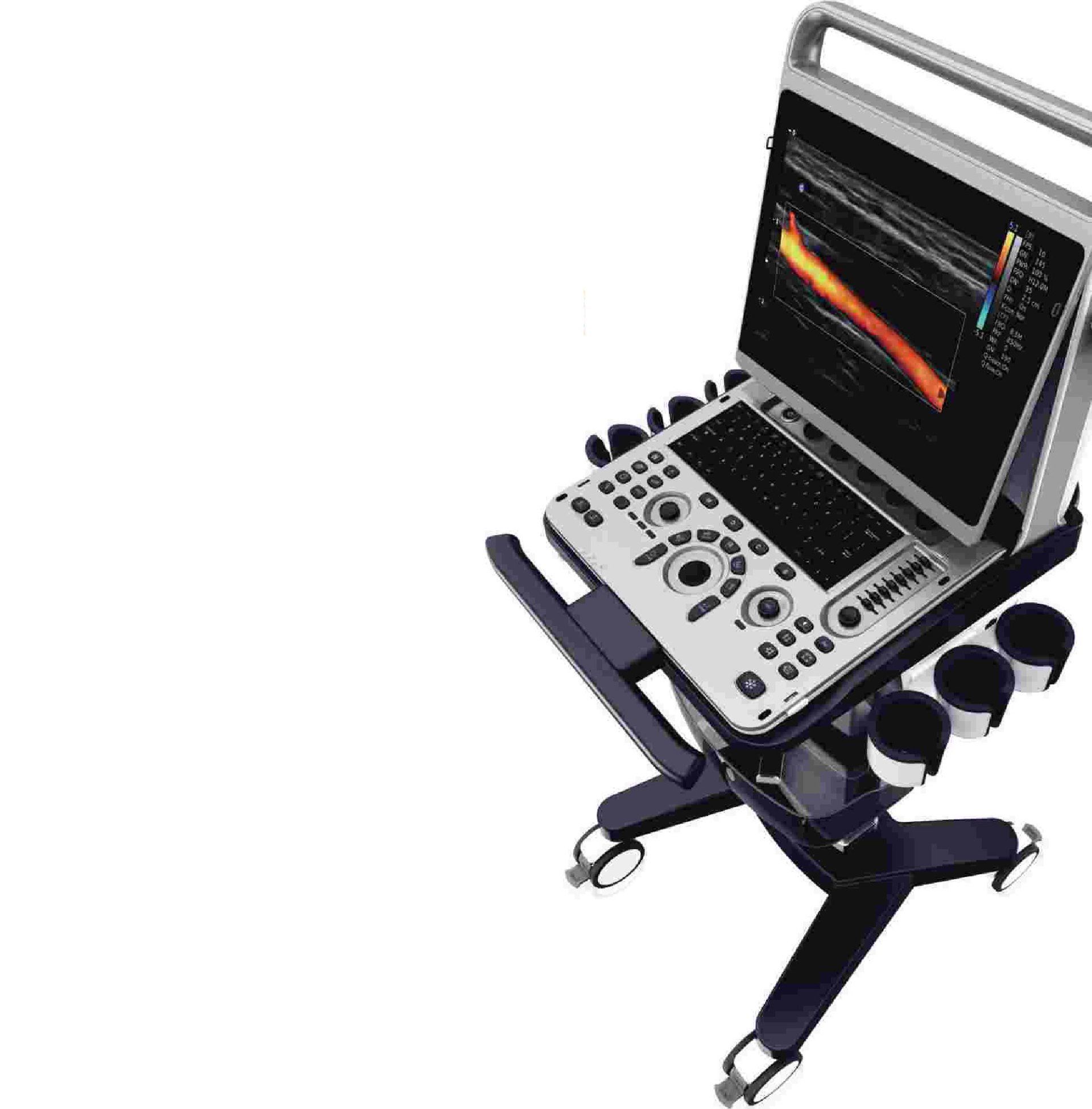
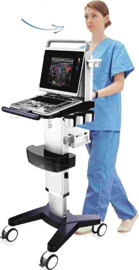
Ergonomic Design
• lndependent angle lS"LED ( Oº - 30º tilted )
• Lightweight ( 7.SKg / 16.Slbs)
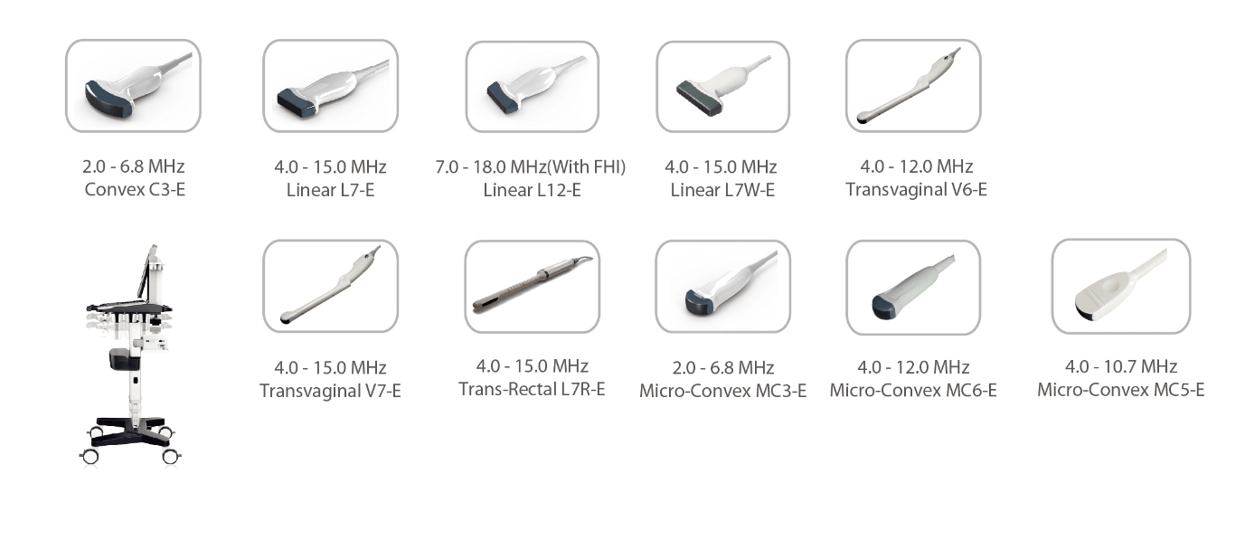
• Dual transducer ports ( Built-in )
• Probe holders
• Removable battery, 120 minutes in active mode
• lndependent angle lS"LED ( Oº - 30º tilted )
• Lightweight ( 7.SKg / 16.Slbs)
• Dual transducer ports ( Built-in )
• Probe holders
• Removable battery, 120 minutes in active mode

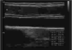
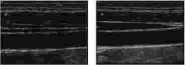
Auto IMT
Automatically traces the intima, and measures the thickness of the intima. This allows you to measure the intima faster, more easily and more accurately
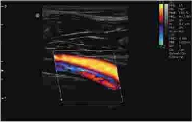
Up to 18MHz High
Frequency Linear Probe
Our high frequency linear probe provides unparalleled detail resolution and superior contrast resolution with up to 18 MHz imaging frequency.
Q-image
These innovative algorithms have strengthened the image enhancement results significantly. Advanced chipset is used to ensure fast frame rate.
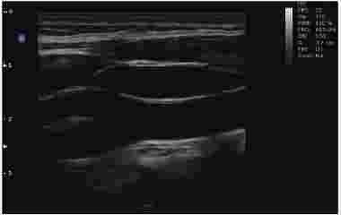
Super Needle
With Super Needle, clinicians can see needle inside tissue more clearly during medical procedures. Needle angle up to ±30º

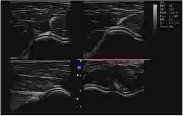
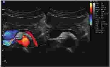
CCA, ICA, ECA, 8 Mode
Popliteal Artery and Vein
Fingertips Vessle, C Mode
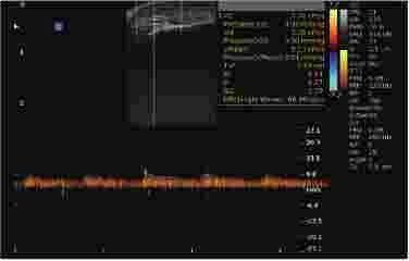
Fingertips Vessle, PW Mode
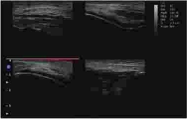
Knee , 48 Mode
Elbow Joint, 48 Mode
General lmaging
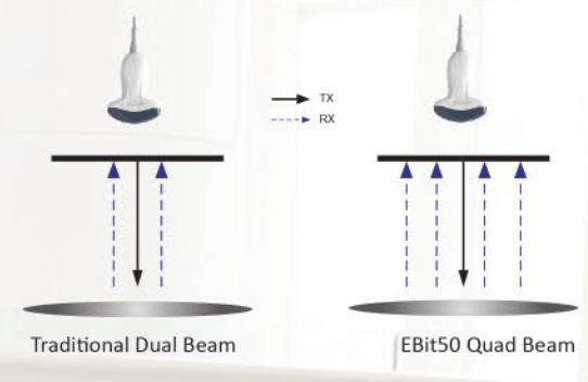
Q-beam
- Compared to the traditional dual-beam former on most ultrasound machines, the EBitSO uses quad-beam technology for ultrasound signal receiving.
- Increasing image resolution and generating more accurate images.
- Produces higher frame rates, ensuring better diagnostic confidence and efficiency, especially for moving organs.Treatment times are quick and efficient
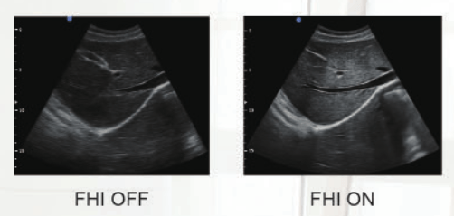
FHI
- FHI is an innovative harmonic imaging technology that uses multiple transmission and receiving methods based on the patients' size and weight. This allows the EBit to maintain image resolution when imaging larger patients.
- Traditional Tissue Harmonics and Phased Harmonics compromise image quality and resolution when penetration is increased.
- Chison's FHI technology greatly improves diagnostic abilities and clinical confidence in larger, difficult-to-image patients.
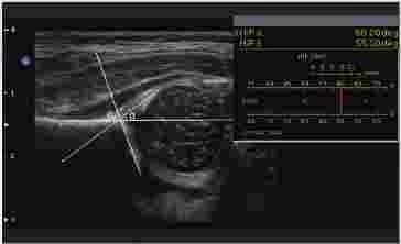
HIP Graf
Gallbladder stone, B Mode
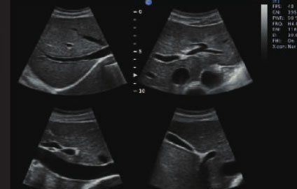
Abdomen, 48 Mode
Pancreas, B/BC Mode

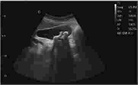
Umbilical cord, C Mode
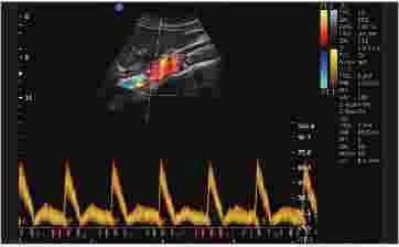
Aorta Artery, PW Mode
Specifications
- 8, 28, 48, 8 / M
- CFM ePW
- PD, DPD(Direction Power Doppler)
- Duplex, Triplex
- Trapezoidal
- Chroma 8/ M / PW
- Super Needle ( option )
- 20 steer
- Auto IMT
- HIP Graf
- DICOM ( option )
Comprehensive Applications
- OB/GYN
- Urology
- Pediatric
- Radiology
- Interna! Medicine
- Small Parts
- General lmaging
- Vascular
- lntensive Care
- Emergency
- MSK
lmage Processing Technologies
- FHI
- Q-beam
- Q-flow
- Q-image
- X-contrast
- SRA
- Compound Image
Accessories
- Footswitch
- Trolley
- Suitcase
- Video Printer
- PC Printer